
Evaluation of coronary arteries stenosis by computed tomography angiography in district Faisalabad
Article Sidebar
Main Article Content
Abstract
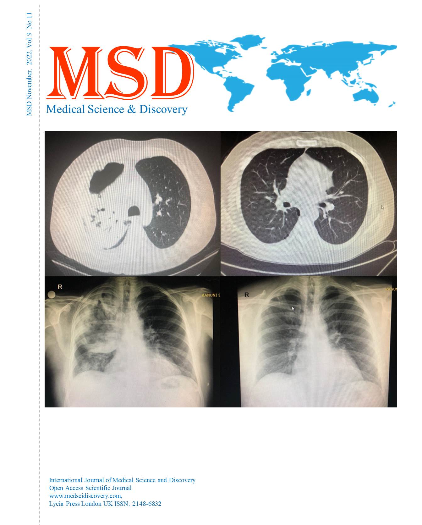
Objective: This study was conducted to investigate the accuracy and precision of computed tomography angiography for assessing significant coronary artery stenosis in district Faisalabad.
Materials and Methods: The data was collected from the Radiology department of Shifa International hospital Faisalabad. Sixty patients (40-80 years of age) were assessed through computed tomography to evaluate coronary heart disease. Data was collected by using a close-ended self-modified questionnaire and analyzed by SPSS V22.
Results: The findings of this study showed that the incidence of coronary heart disease patients was 34 (56.67%) out of 60. Among the affected patients, 39 (65%) were male, and 21 (35%) were female. Based on the evaluation, most of the affected patients were under (50-60) years of age. The percentage of coronary arteries stenosis varies as the left anterior-descending artery LAD had higher stenosis (41.67%) among others; left main LM (16.67%), left-circumflex artery LCX (20%), and right-coronary artery RCA (21.67%) in all affected patients.
Conclusion: It was presumed that computed tomography angiography precisely distinguishes the presence and finding of coronary stenosis and was additionally pronounced the best quality level. Based on coronary arteries, stenosis of LAD was the most commonly reported in diseased patients. Other arteries stenosis can also increase the risk of CHD in patients.
Downloads
Article Details

This work is licensed under a Creative Commons Attribution-NonCommercial 4.0 International License.
Accepted 2022-11-16
Published 2022-11-19
References
Sharma A, Arbab-Zadeh A. Assessment of coronary heart disease by CT angiography: current and evolving applications. Journal of Nuclear Cardiology. 2012 Aug;19(4):796-806.
Sanchis-Gomar F, Perez-Quilis C, Leischik R, Lucia A. Epidemiology of coronary heart disease and acute coronary syndrome. Annals of translational medicine. 2016 Jul;4(13).
Schoepf UJ, Becker CR, Ohnesorge BM, Yucel EK. CT of coronary artery disease. Radiology. 2004 Jul;232(1):18-37.
Chilton RJ. Pathophysiology of coronary heart disease: a brief review. Journal of Osteopathic Medicine. 2004 Sep 1;104(s9):5-8.
Koba S, Hirano T, Kondo T, Shibata M, Suzuki H, Murakami M, Geshi E, Katagiri T. Significance of small dense low-density lipoproteins and other risk factors in patients with various types of coronary heart disease. American Heart Journal. 2002 Dec 1;144(6):1026-35.
Stone GW, Maehara A, Lansky AJ, De Bruyne B, Cristea E, Mintz GS, Mehran R, McPherson J, Farhat N, Marso SP, Parise H. A prospective natural-history study of coronary atherosclerosis. New England Journal of Medicine. 2011 Jan 20;364(3):226-35.
Bowman AW, Kantor B, Gerber TC. Coronary computed tomographic angiography: current role in the diagnosis and management of coronary artery disease. Polskie Archiwum Medycyny Wewnetrznej. 2009 Jun;119(6):381.
Anderson JL, Morrow DA. Acute myocardial infarction. New England Journal of Medicine. 2017 May 25;376(21):2053-64.
Gould KL. Quantification of coronary artery stenosis in vivo. Circulation research. 1985 Sep;57(3):341-53.
El-Menyar AA, Al Suwaidi J, Holmes Jr DR. Left main coronary artery stenosis: state-of-the-art. Current problems in cardiology. 2007 Mar 1;32(3):103-93.
El-Menyar AA, Al Suwaidi J, Holmes Jr DR. Left main coronary artery stenosis: state-of-the-art. Current problems in cardiology. 2007 Mar 1;32(3):103-93.
Lloyd-Jones DM, Larson MG, Beiser A, Levy D. Lifetime risk of developing coronary heart disease. The Lancet. 1999 Jan 9;353(9147):89-92.
Zerwic JJ, King KB, Wlasowicz GS. Perceptions of patients with cardiovascular disease about the causes of coronary artery disease. Heart & Lung. 1997 Mar 1;26(2):92-8.
Libby P, Theroux P. Pathophysiology of coronary artery disease. Circulation. 2005 Jun 28;111(25):3481-8.
Beck CS, Leighninger DS. Operations for coronary artery disease. Journal of the American Medical Association. 1954 Nov 27;156(13):1226-33..
Mowatt G, Cook JA, Hillis GS, Walker S, Fraser C, Jia X, Waugh N. 64-Slice computed tomography angiography in the diagnosis and assessment of coronary artery disease: systematic review and meta-analysis. Heart. 2008 Nov 1;94(11):1386-93.
Ostrom MP, Gopal A, Ahmadi N, Nasir K, Yang E, Kakadiaris I, Flores F, Mao SS, Budoff MJ. Mortality incidence and the severity of coronary atherosclerosis assessed by computed tomography angiography. Journal of the American College of Cardiology. 2008 Oct 14;52(16):1335-43.
Chow BJ, Wells GA, Chen L, Yam Y, Galiwango P, Abraham A, Sheth T, Dennie C, Beanlands RS, Ruddy TD. Prognostic value of 64-slice cardiac computed tomography: severity of coronary artery disease, coronary atherosclerosis, and left ventricular ejection fraction. Journal of the American College of Cardiology. 2010 Mar 9;55(10):1017-28.
von Ballmoos MW, Haring B, Juillerat P, Alkadhi H. Meta-analysis: diagnostic performance of low-radiation-dose coronary computed tomography angiography. Annals of internal medicine. 2011 Mar 15;154(6):413-20.
Chow BJ, Small G, Yam Y, Chen L, Achenbach S, Al-Mallah M, Berman DS, Budoff MJ, Cademartiri F, Callister TQ, Chang HJ. Incremental prognostic value of cardiac computed tomography in coronary artery disease using CONFIRM: COroNary computed tomography angiography evaluation for clinical outcomes: an InteRnational Multicenter registry. Circulation: Cardiovascular Imaging. 2011 Sep;4(5):463-72.
Schoepf UJ, Zwerner PL, Savino G, Herzog C, Kerl JM, Costello P. Coronary CT angiography. Radiology. 2007 Jul;244(1):48-63.
Gilard M, Le Gal G, Cornily JC, Vinsonneau U, Joret C, Pennec PY, Mansourati J, Boschat J. Midterm prognosis of patients with suspected coronary artery disease and normal multislice computed tomographic findings: a prospective management outcome study. Archives of internal medicine. 2007 Aug 13;167(15):1686-9.
Bluemke DA, Achenbach S, Budoff M, Gerber TC, Gersh B, Hillis LD, Hundley WG, Manning WJ, Printz BF, Stuber M, Woodard PK. Noninvasive coronary artery imaging: magnetic resonance angiography and multidetector computed tomography angiography: a scientific statement from the american heart association committee on cardiovascular imaging and intervention of the council on cardiovascular radiology and intervention, and the councils on clinical cardiology and cardiovascular disease in the young. Circulation. 2008 Jul 29;118(5):586-606.
Rubinshtein R, Halon DA, Gaspar T, Jaffe R, Karkabi B, Flugelman MY, Kogan A, Shapira R, Peled N, Lewis BS. Usefulness of 64-slice cardiac computed tomographic angiography for diagnosing acute coronary syndromes and predicting clinical outcome in emergency department patients with chest pain of uncertain origin. Circulation. 2007 Apr 3;115(13):1762-8.
Leber AW, Knez A, von Ziegler F, Becker A, Nikolaou K, Paul S, Wintersperger B, Reiser M, Becker CR, Steinbeck G, Boekstegers P. Quantification of obstructive and nonobstructive coronary lesions by 64-slice computed tomography: a comparative study with quantitative coronary angiography and intravascular ultrasound. Journal of the American College of Cardiology. 2005 Jul 5;46(1):147-54.
Raff GL, Gallagher MJ, O’Neill WW, Goldstein JA. Diagnostic accuracy of noninvasive coronary angiography using 64-slice spiral computed tomography. Journal of the American College of Cardiology. 2005 Aug 2;46(3):552-7.
Sato A, Nozato T, Hikita H, Seo Y, Ishizu T, Murakoshi N, Sakai S, Watanabe S, Aonuma K. Coronary Artery Spatial Distribution, Morphology, and Composition of Non-culprit Vulnerable Plaques by 64-slice Computed Tomography Angiography in Patients With Acute Myocardial Infarction.
Budoff MJ, Rasouli ML, Shavelle DM, Gopal A, Gul KM, Mao SS, Liu SH, McKay CR. Cardiac CT angiography (CTA) and nuclear myocardial perfusion imaging (MPI)—a comparison in detecting significant coronary artery disease. Academic radiology. 2007 Mar 1;14(3):252-7.
Nissen SE. Limitations of computed tomography coronary angiography. Journal of the American College of Cardiology. 2008 Dec 16;52(25):2145-7.
Schuijf JD, Achenbach S, de Feyter PJ, Bax JJ. Current applications and limitations of coronary computed tomography angiography in stable coronary artery disease. Heart. 2011 Feb 15;97(4):330-7.
Sun Z, Jiang W. Diagnostic value of multislice computed tomography angiography in coronary artery disease: a meta-analysis. European journal of radiology. 2006 Nov 1;60(2):279-86.
Sun Z, Choo GH, Ng KH. Coronary CT angiography: current status and continuing challenges. The British journal of radiology. 2012 May;85(1013):495-510.
Arbab-Zadeh A, Miller JM, Rochitte CE, Dewey M, Niinuma H, Gottlieb I, Paul N, Clouse ME, Shapiro EP, Hoe J, Lardo AC. Diagnostic accuracy of computed tomography coronary angiography according to pre-test probability of coronary artery disease and severity of coronary arterial calcification: the CORE-64 (Coronary Artery Evaluation Using 64-Row Multidetector Computed Tomography Angiography) international multicenter study. Journal of the American College of Cardiology. 2012 Jan 24;59(4):379-87.
Busch JL, Alessio AM, Caldwell JH, Gupta M, Mao S, Kadakia J, Shuman W, Budoff MJ, Branch KR. Myocardial hypo-enhancement on resting computed tomography angiography images accurately identifies myocardial hypoperfusion. Journal of cardiovascular computed tomography. 2011 Nov 1;5(6):412-20.
Min JK, Dunning A, Lin FY, Achenbach S, Al-Mallah M, Budoff MJ, Cademartiri F, Callister TQ, Chang HJ, Cheng V, Chinnaiyan K. Age-and sex-related differences in all-cause mortality risk based on coronary computed tomography angiography findings: results from the International Multicenter CONFIRM (Coronary CT Angiography Evaluation for Clinical Outcomes: An International Multicenter Registry) of 23,854 patients without known coronary artery disease. Journal of the American College of Cardiology. 2011 Aug 16;58(8):849-60.
Motoyama S, Sarai M, Harigaya H, Anno H, Inoue K, Hara T, Naruse H, Ishii J, Hishida H, Wong ND, Virmani R. Computed tomographic angiography characteristics of atherosclerotic plaques subsequently resulting in acute coronary syndrome. Journal of the American College of Cardiology. 2009 Jun 30;54(1):49-57.
Gaemperli O, Schepis T, Valenta I, Koepfli P, Husmann L, Scheffel H, Leschka S, Eberli FR, Luscher TF, Alkadhi H, Kaufmann PA. Functionally relevant coronary artery disease: comparison of 64-section CT angiography with myocardial perfusion SPECT. Radiology. 2008;248(2):414-23.
Meijboom WB, van Mieghem CA, Mollet NR, Pugliese F, Weustink AC, Van Pelt N, Cademartiri F, Nieman K, Boersma E, de Jaegere P, Krestin GP. 64-slice computed tomography coronary angiography in patients with high, intermediate, or low pretest probability of significant coronary artery disease. Journal of the American College of Cardiology. 2007 Oct 9;50(15):1469-75.
Leschka S, Alkadhi H, Plass A, Desbiolles L, Grünenfelder J, Marincek B, Wildermuth S. Accuracy of MSCT coronary angiography with 64-slice technology: first experience. European heart journal. 2005 Aug 1;26(15):1482-7.